European Conference on Embolotherapy
ET countries

June 1-4 | Vienna, Austria

Mastering embolization

June 1-4 | Vienna, Austria

Mastering embolization

June 1-4 | Vienna, Austria

Mastering embolization

June 1-4 | Vienna, Austria

June 1-4 | Vienna, Austria

Slider
ProgrammeSneak peeks overviewEndoleaks – when to intervene?

Endoleaks – when to intervene?


Three reasons why you cannot miss my lecture

  1. You will learn about optimal imaging and follow-up strategies post EVAR
  2. You will understand the indications for treating different types of endoleak
  3. You will learn about the available treatment options

Dr. Joo-Young Chun
Speaker bio
 

Add this session to your calendar!

Why do endoleaks matter?

Endoleaks are the most common complication after EVAR and may compromise the durability of aortic repair. There are five types of endoleak based on their anatomical site and aetiology (table 1) and their management depends on the type and associated risk of sac rupture. Some types are associated with ongoing risk of aneurysm rupture and require long-term surveillance and secondary interventions. Early detection and correct classification of endoleaks are essential to guide decision-making around potential re-intervention. The majority of endoleaks that require treatment can be managed using endovascular techniques.

Endoleak typeLocation of leakIncidence (%)
Type IAttachment sites2-10
AProximal end
BDistal end
CIliac occluder
Type IIRetrograde flow through patient aortic side branches8-29
ASingle vessel
BMultiple vessels
Type IIIMechanical failure1-5
AModular disconnection
BFabric tear
CJunctional separation (fenestration, branch, visceral stent)
Type IVGraft porosity<1
Type VAneurysm sac enlargement without visualised endoleak2-3

Table 1: Endoleak types and their reported incidence

Imaging surveillance after EVAR

Imaging surveillance is necessary in all patients who undergo EVAR to identify complications including endoleak, sac growth, and stent graft migration. A typical surveillance protocol includes CT at 1 month and at 12 months post EVAR. If the 1-month scan shows type 1 or 3 endoleak or unexplained sac increase, an additional scan at 6 months is advisable. If the 12-month scan shows stable sac size with either type 2 or no endoleak, further annual follow-up with duplex is deemed safe. Detection of a new endoleak or sac expansion on duplex ultrasound should prompt further CT evaluation.

Contrast-enhanced US is in conjunction with CTA to assess the flow dynamics of an endoleak, or in unexplained sac expansion with a negative CTA where US microbubbles may demonstrate a slow type 2 endoleak. Similarly, MRI may be used to look for occult endoleaks in cases of unexplained sac expansion although susceptibility artefact from metallic stent grafts and coils may limit image interpretation.

Conventional catheter angiography +/- cone-beam CT may be used as a problem-solving tool when the type and source of an endoleak is inconclusive, for example a type 1 endoleak at the seal zones may be confused with a type 2 endoleak. Rotational catheter angiography with cone-beam CT can be useful in detecting slow-flow type 2 endoleaks in cases of unexplained sac expansion.

Indications for treatment

Type 1 endoleaks represent failure of proximal or distal seal and are associated with elevated sac pressure and an ongoing risk of aneurysm expansion and rupture. They warrant prompt treatment upon detection. Treatment options include aortic cuff, fenestrated cuff, chimney graft, distal stent graft extension and embolization.

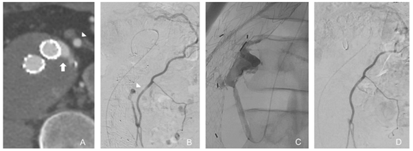
Type 2 endoleaks are caused by retrograde blood flow from aortic branches and are inherently low flow and often transient. They usually involve the IMA and lumbar arteries although accessory renal and internal iliac artery branches may also be involved. They are managed conservatively unless associated with sac expansion. The mainstay of treatment is embolization with a range of embolic agents via multiple routes including transarterial (Figure 1), direct sac puncture or transcaval approaches.

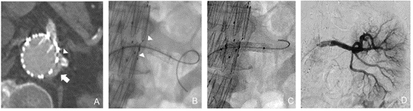
Type 3 endoleaks arise from a structural defect or modular disconnection of stent graft components. This leads to an increase in sac pressure and ongoing risk of aneurysm rupture. They warrant prompt treatment upon detection, usually with additional stent grafts to reline a fabric tear or bridge across a gap (Figure 2).

Type 5 endoleaks represent cases of unexplained sac expansion without an identifiable endoleak. These are challenging to manage and warrant further imaging with multiple modalities to exclude an occult endoleak in the first instance.

Figure 1: Transarterial embolization of type 2 endoleak. (A) Axial CT shows type 2 endoleak (arrow) in anterior sac close to the IMA (arrowhead). (B) Angiogram from middle colic branch of SMA shows hypertrophied arc of riolan, IMA and endoleak cavity (arrowhead). (C) Embolisation with a liquid embolic agent via microcatheter. (D) Completion angiogram shows no further endoleak opacification
Figure 2: Type 3 endoleak in a fenestrated EVAR. (A) Axial CT shows junctional separation between the main body and fenestrated left renal stent (arrowhead) resulting in type 3 endoleak (arrow). (B) Fluoroscopic image shows a visible gap between the two stent graft components (arrow heads). (C) Additional renal stent graft deployed to bridge the gap. (D) No residual endoleak on completion angiogram.


Joo-Young Chun

St. George's University Hospital<, London/GB


Dr Joo-Young Chun is a consultant interventional radiologist at St George’s University Hospital with a special interest in vascular intervention. Her research interests include management of endoleak, haemoptysis, peripheral vascular disease, and major trauma. She is an active member of CIRSE and serves as editor-in-chief of A-type questions on the EBIR Examination Council and as CVIR Endovascular Associate Editor. She is also a member of the Clinical Services in IR Task Force and the CIRSE 2024 Scientific Programme Committee. She was a member of the Standards of Practice Committee and led the publication of two SOP documents; Management of Endoleaks and Conducting Morbidity & Mortality meetings.


References

  1. Chun JY, de Haan M, Maleux G, Osman A, Cannavale A, Morgan R. CIRSE Standards of Practice on management of endoleaks following endovascular aneurysm repair. Cardiovasc Intervent Radiol (2024) 47:161–176
  2. van Marrewijk C, Buth J, Harris PL, Norgren L, Nevelsteen A, Wyatt MG. Significance of endoleaks after endovascular repair of abdominal aortic aneurysms: The EUROSTAR experience. J Vasc Surg. 2002;35(3):461–73
  3. Wanhainen A, Verzini F, Van Herzeele I, Allaire E, Bown M et al. European Society for Vascular Surgery (ESVS) 2019 Clinical Practice Guidelines on the Management of Abdominal Aorto-liac Artery Aneurysms. Eur J Vasc Endovasc Surg. 2019;57(1):8–93
  4. Ameli-Renani S, Pavlidis V, Morgan RA. Secondary endoleak management following TEVAR and EVAR. Cardiovasc Intervent Radiol. 2020;43(12):1839–54
  5. Cannavale A, Lucatelli P, Corona M, Nardis P, Basilico F, DeRubeis G, Santoni M, Catalano C, Bezzi M. Evolving concepts and management of endoleaks after endovascular aneurysm repair: where do we stand in 2019? Clin Radiol. 2020;75(3):169–78.
  6. Chung R, Morgan RA. Type 2 endoleaks post-EVAR: current evidence for rupture risk, intervention, and outcomes of treatment. Cardiovasc Intervent Radiol. 2015;38(3):507–2